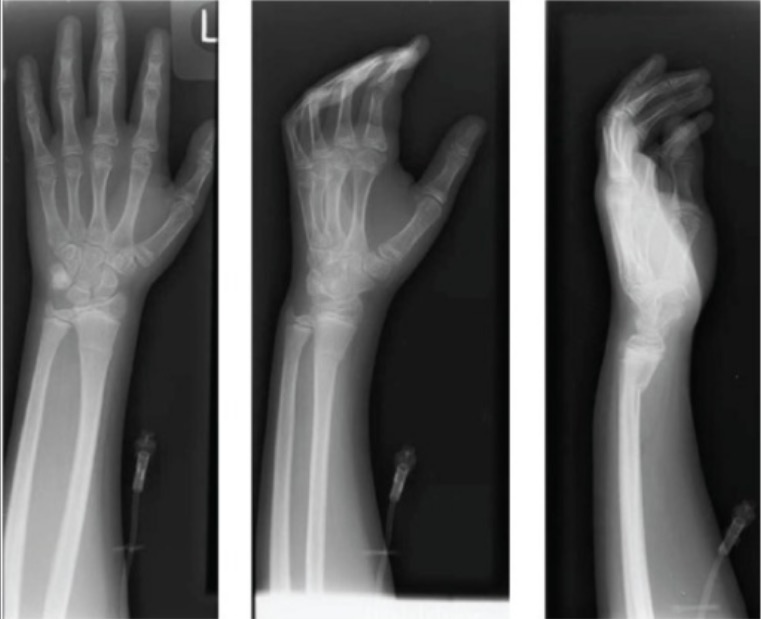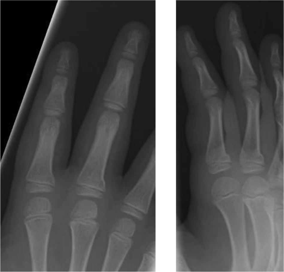
DL models commonly build upon large training image sets for robust outcomes ( 7, 8), often containing thousands, hundred-thousands, millions or more different samples. With few exceptions, they fall into the domain of deep learning (DL) through convolutional neural networks (CNN) ( 5, 6). Radiological AI models usually originate from annotated image data, also known as supervised AI ( 4).

Prior studies demonstrated that Artificial Intelligence (AI) is able to successfully detect fractures in radiographs ( 1– 3). The resulting images aggregate into a complete examination for interpretation by emergency physicians or radiologists. Sometimes, additional projections are performed. Standard DR procedures include two orthogonally-oriented projections of the wrist joint and the adjacent structures. Injuries around the wrist are typically examined by digital radiography (DR). In adults and children, the distal radius is the most common site for fractures. Built into the radiography workflow, such an algorithm could contribute to radiation hygiene and patient comfort. Following training on larger data sets CNNs might be able to effectively rule out the presence of a distal radius fracture, enabling to consider foregoing the yet inevitable lateral projection in children. CNN predictions reached area under the curves (AUCs) up to 98%, consistently exceeding human expert ratings (mean AUC 93.5%, 95% CI 89.9%–97.2%). In total, 200 previously unseen images (100 per class) served as test set. The task was to classify images into fractured and non-fractured. We trained three different state-of-the-art convolutional neural networks (CNNs) on a dataset of 2,474 images: 1,237 images were posteroanterior (PA) pediatric wrist radiographs containing isolated distal radius torus fractures, and 1,237 images were normal controls without fractures.

However, there is cautious hope that computer vision could enable breaking with this tradition in minor injuries, clinically lacking malalignment. It is an indisputable dogma in extremity radiography to acquire x-ray studies in at least two complementary projections, which is also true for distal radius fractures in children.


 0 kommentar(er)
